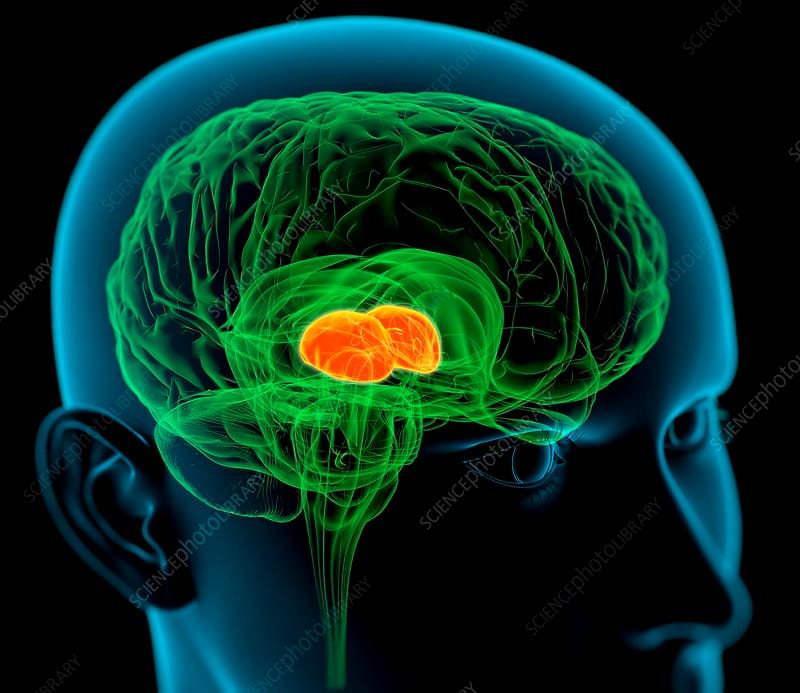
Thalamus - Relay for CNS signal processing

Every major city has a central hub through which its citizens can access public transportation to get to their desired destination. In the case of the human body, the nervous system can be divided into the major road ways (nerves) that carry individuals (impulses) to and from the big city (the brain).
What is the thalamus and what does it do?
The thalamus is often described as a relay station. This is because almost all sensory information (except for smell) that proceeds to the cortex first stops in the thalamus before being sent on to its destination. The thalamus is subdivided into a number of nuclei that possess functional specializations for dealing with particular types of information. Sensory information thus travels to the thalamus and is routed to a nucleus tailored to dealing with that type of sensory data. Then, the information is sent from that nucleus to the appropriate area in the cortex where it is further processed.
For example, visual information from your retina travels to the lateral geniculate nucleus of the thalamus, which is specialized to handle visual information, before being sent on to the primary visual cortex (the main area for visual processing in the brain). A similar pathway through the thalamus can be delineated for all sensory information except smell. In fact, the majority of all the signals (not just sensory) that pass to the cortex first pass through the thalamus.
Gross structure
The thalamus is made up of two symmetrical structures formed from the diencephalon. Each half of the thalamus is elongated along the anteroposterior axis giving it an ovoid appearance. It is narrowest at the anterior end and widest at the posterior part.
The thalami are made up of grey matter that is partitioned by a “Y” shaped white matter structure known as the internal medullary lamina.
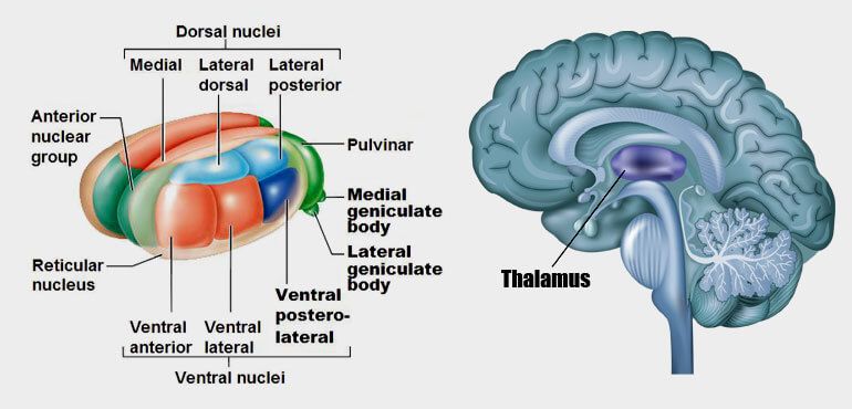
As a result of the location of the internal medullary lamina, each thalamus is divided into roughly three main parts: the anterior, medial and lateral thalamus. The anterior part lies between the short limbs of the internal medullary lamina, while the medial and lateral parts lie on the respective side of the main stem of the “Y”. The left thalamus communicates with the right thalamus by way of the interthalamic adhesion.
The overall appearance of the thalamus may seem unremarkable with the exception of two protuberances on the posteroventral surface. These are the medial and lateral geniculate bodies, which are responsible for the processing of auditory and visual sensory inputs, respectively. Furthermore, the thalami are each surrounded two layers of white matter.
Dorsally, it is covered by a layer known as the stratum zonale; while laterally, it is covered by the external medullary lamina, which separates the lateral and ventral thalamus from the thalamic reticular nucleus and the sub thalamus.
Relations
Anterior
Its anterior pole forms the posterior wall of the inter ventricular foramen of Monro, which permits communication between the lateral and the third ventricles. Additionally, there are five veins that coalesce to form the internal cerebral vein at the anterior end of the thalamus. These vessels can be remembered as STACS (superior striate, thalamostriate, anterior terminal, choroidal and septal veins).
Medial
Furthermore, the medial wall of each thalamus forms the lateral walls of the aforementioned third ventricle.
Dorsal
The dorsal surface is in close relation to the stria terminalis, the choroid plexus of the third ventricle and the body of the fornix. The internal cerebral vein courses along the dorsomedial length of the thalamus, while the superior thalamostriate vein runs along the dorsolateral surface.
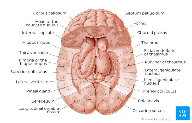
Superficial to the stratum zonale of the thalamus is the caudate nucleus. The head of the caudate nucleus lies anterosuperiorly to the thalamus with the body travelling superior and laterally to the body of the thalamus. Lateral to the external medullary lamina is the reticular nucleus, then the posterior limb of the internal capsule. Numerous neuronal tracts travel through the different limbs of the internal capsule to synapse in the thalamus.
Furthermore, the internal capsule also separates the thalamus from the globus pallidus and putamen (collectively known as the lentiform nucleus).
Posterior
The posterior most aspect of the thalamus is known as the pulvinar. Each pulvinar is lateral to the pineal gland, the Habenular and posterior commissures, posterolateral to the corpora quadrigemini (superior and inferior colliculi), and superior to the medial and lateral geniculate bodies. Additionally, the posterior thalamus is deep to the splenium of the corpus callosum.
Inferior
There are two major structures lying inferiorly to the thalamus. Anteroinferiorly is the hypothalamus, caudal to the hypothalamic sulcus. Directly inferior to the thalamus is the cerebral peduncle and the cerebral aqueduct of Sylvius. Also note that the inferior surface of the thalamus is continuous with the tegmentum of the (floor of the midbrain between the cerebral peduncle and the quadrigeminal plate).
Source: interactive-biology, kenhub.com