Advanced Review Labelling of neuronal receptors and transporters with quantum dots.

The ability to efficiently visualize protein targets in cells is a fundamental goal in biological research. Recently, quantum dots (QDots) have emerged as a powerful class of fluorescent probes for labelling membrane proteins in living cells because of breakthrough advances in QDot surface chemistry and bio-functionalization strategies. This review discusses the increasing use of QDots for fluorescence imaging of neuronal receptors and transporters. The readers are briefly introduced to QDot structure, photophysical properties, and common synthetic routes toward the generation of water-soluble QDots. The following section highlights several reports of QDot application that seek to unravel molecular aspects of neuronal receptor and transporter regulation and trafficking. This article is closed with a prospectus of the future of derivatized QDots in neurobiological and pharmacological research.
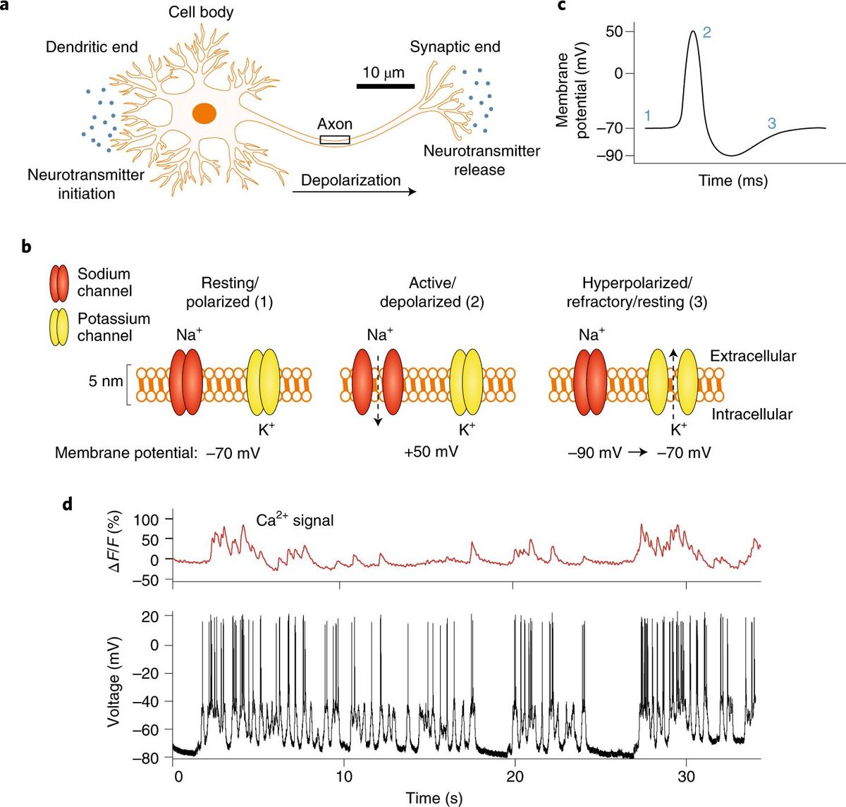
Since the pioneering neuroanatomical work of Santiago Ramon y Cajal in the early 20th century, the need to visualize dynamic communication within neuronal networks has been a challenge for modern neuroscience.1 Synaptic transmission, and hence central nervous system excitability, are modulated in a tightly regulated manner by a myriad of neuronal membrane receptors and transporters that rely on specific ligand binding and transport and the initiation of intracellular signalling cascades. Despite extensive biochemical and genetic analyses, mechanisms that regulate synaptic receptor and transporter activity, trafficking, and localization continue to challenge neuroscientists.
These considerations have driven our desire to combine recently developed receptor and transporter labelling methods with advanced fluorescence imaging tools to dissect mechanisms of synaptic protein trafficking and signalling at the single molecule level. Fluorescent labelling techniques commonly used to interrogate cellular processes can be classified into two broad categories: (1) the construction and expression of fluorescent fusion proteins such as green fluorescent protein (GFP) and (2) the chemical methods of fluorescent labelling. With the rapid evolution of fluorescent protein technology, GFP-based labelling methods have quickly found widespread use as the proteins produced bypass many time-consuming probe preparation steps and assure that labeling is restricted to the desired proteins.
Unfortunately, there are certain limitations associated with the GFP fusion approach: (1) incompatibility of GFP construct transfection with endogenous expression systems; (2) failure of GFP-tagged proteins to localize properly; and (3) differences in the activity and signaling of GFP-tagged proteins compared to their wild-type counterparts. As a result, alternative fluorescence labelling strategies have emerged for the visualization of cellular proteins, particularly those systems that are incompatible with the GFP based methodology. However, as chemical labelling techniques typically utilize conventional fluorescent dyes, live-cell imaging is often hampered by the low photostability and brightness, narrow Stokes shift, and relatively broad emission spectra of these labels. To address these shortcomings, researchers have developed fluorescent nanomaterials with significantly improved optical properties for biological imaging.
Semiconductor nanocrystals, also known as quantum dots (QDots), are one such class of fluorescent nanomaterials that overcome instability issues associated with conventional fluorescent dyes and support versatile protein labelling via their surface
functionalization with various targeting probes. More importantly, QDots are much better suited to advanced fluorescence imaging approaches that pursue live-cell, multicolour detection, in vivo animal imaging,10–12 and single-molecule tracking. Here, an overview of recent advances in QDot synthesis and the conjugation of biological probes to the QDot surface is provided. In addition, several reports of QDot application that seek to unravel molecular aspects of neuronal receptor and transporter regulation and trafficking are highlighted. This article is closed with a prospectus of the future of derivatized QDots in neurobiological and pharmacological research. the as-synthesized QDot is composed of a CdSe core capped with a layer of hydrophobic ligands.
These ‘bare’, water-insoluble QDots cannot be immediately used in an aqueous, biological environment; moreover, these QDots are susceptible to degradation/aggregation and suffer from relatively low fluorescence quantum yields as a result of the nonradiative recombination processes occurring at the core surface. To eliminate these problems, the CdSe core is commonly passivated with a shell of a wider bandgap inorganic material (ZnS) to increase its stability and optimize optical properties.16,18 Thus, the term ‘quantum dot’ usually refers to a CdSe/ZnS core/shell structure.
What Is a QDot?
QDots are nanometer-sized crystals that are composed of several hundreds to thousands of atoms of periodic group II/VI (CdSe, CdTe, CdS), IV/VI (PbS, PbSe), III/V (InP), or ternary (InGaP) semiconductor materials, with CdSe being one of the most commonly encountered materials.
Full study: source